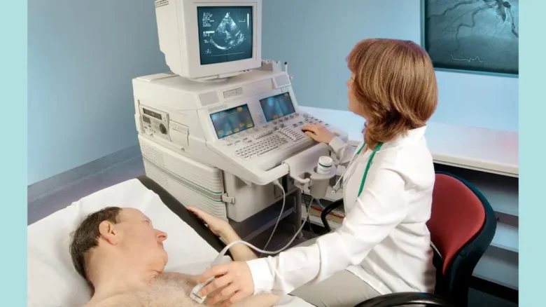
In recent times, issues with heart health have arisen and become areas of concern for many. Out of the many options available for a cardiologist, echocardiography is one of the best options available for diagnosing and managing heart conditions. It is a completely non-invasive procedure wherein high-frequency sound waves are used to form images of the heart, allowing for a comprehensive assessment of its anatomy and physiology. We will see how these technologies are impacting the management of heart health and what their future looks like in the heart troubleshooting space in this article.
Echocardiography and its Importance in Maintaining Heart Health:
The technology of echocardiography has been amongst the very first cardiac diagnostic tools to have been invented. Rather, underdeveloped technologies were used to allow for seeing a part of the heart and its rotation and how effective it is at pumping blood while identifying any inconsistencies. The patients far benefit now as using the technology allows for the identification of valve disease, cardiomyopathy, and even arrhythmias without having to deliver a surgical procedure.
Normally, chest echocardiography is done by placing a transducer on the chest. This transducer produces sound waves directed at the heart, which reflects them. The reflected sound waves or echoes are then converted into visual images, which help doctors observe the patient’s heart by viewing its valves and other structures of interest.
One of the crucial facilities in the emergency department and as well in the routine assessment is echocardiography. It is being applied in hospitals across the globe in order to assist in making management choices and assessing disease progression. Nowadays, it is an essential part of clinical cardiology due to its non-invasive nature and adequate diagnosis.
Historical Echocardiographic Approaches:
There are several techniques, which include M-mode, 2D, and Doppler echocardiography, which are classified under echocardiography. These techniques are uniquely different in their approach and offer added value in appreciating heart function in every dimension.
M-mode echocardiography is used to evaluate ejection fraction and visual assessment of the left ventricle contour as well as in the assessment in the event of a dummy of the right heart, provides a vertical one-dimensional ultrasound image of the heart, and is components often used to determine the volume size of the heart valves span and thickness of the coral wall ave as well. While it is pretty / slightly of very limited scope, it can still be relied on for measuring accuracy essential for the diagnosis and treatment of patients.
By contrast, 2D echocardiography involves the three-dimensional imaging of the heart. This way, the clinician can see the section of a heart and its movements and structure. This is the most commonly used technique and is used to give context to how the heart operates.
While Doppler echocardiography measured the transvalvular blood flow, it was particularly effective in detecting abnormal blood flow patterns, including those affected by the valves. They may be combined and cardiologists are able to perform a complete examination of the patient’s heart.
Reshaping the Word of Heart: Changes in Echocardiography
The last few years have seen great strides in echocardiography technology, and these advances are set to reshape every aspect of cardiac care. 3-Dimensional (3D) and 4-dimensional (4D) echocardiography assist in enhancing the quality of the heart diagnostic process.
3D echocardiography allows one to visualize the entire heart and all ventricular structures in undoubted detail. It also serves the purpose of modulating a more empowered image of the heart as a whole, meaning this technique is useful for surgical cases where more than one complex heart structure may be present. Unlike the 2-D examination, accurate assessment of the heart chamber configuration includes the quantitative 3D echocardiography method, which features no focal and polar limitations and virtually allows an unrestricted assessment of the heart’s shape.
The fifth dimension is time in 4D echocardiography, which gives a clinician access to the volumes rather than the images. So, now they can see the heart doing its job, even while it is. This is immune to the development and advancement of the healthcare industry. This advancement particularly eliminates the issues related to critical imaging tracking, especially in cases during surgery when the guidance of the decision-making process is involved.
Also, the development of portable echocardiography devices is ensuring that more and more people have access to heart care. Such handheld devices are convenient since they produce quick results and can be used in clinics, ambulances, and even faraway places. This is a truly visionary solution to the changing healthcare needs in the world.
Benefits and Drawbacks of Echocardiography:
There is a lot to gain from the newest echocardiography advancement. They allow better images, which in turn enhance diagnostics as well as the treatment processes. They also augment the early capabilities, for example, the evaluation of valve activity and the evaluation of defects in the heart.
However, the latest techniques are quite restricted in certain areas of the heart. The high price may hinder their use in several areas, most especially those without adequate resources. Furthermore, there is a lack of specialized training required for the proper application of the particular techniques, which may not be present in all healthcare settings.
In spite of the hurdles, the benefits of advanced techniques of echocardiography cannot be underestimated. They enhance the view of the particular or the overall heart status, enabling enhanced diagnosis and treatment of the patients. If current limitations are addressed, these new ideas can change cardiac care dramatically.
The Future of Echocardiography and Its Impact on Heart Health:
Based on previous experiences, the areas where echocardiogram techniques are going to be applied in the future seem to be broader. Advanced versions of the technology will provide useful tools that are related to heart health management in the future. Otherwise, these measures and tools will be useless if they are readily available to patients across the globe.
The use of AI in the field of echocardiography has been estimated to be one of the most striking advances. AI can also accomplish the task of better and faster echocardiographic imaging analysis than human clinicians and therefore avoid the danger of making a mistake in diagnosis. This could facilitate the identification of heart disorders at an early stage to develop relatively customized and effective methods of treatment for this particular source of risk.
Moreover, the creation of portable and simpler-to-operate devices is another developing area of research. These advancements would help reach patients who live in remote or backward areas because there would be a greater scope of echocardiography, which would bring scoring aids closer to the masses. By increasing the availability of such medical diagnosing instruments, we can enhance heart health in the world.
Making Strides in Global Health Care for the Heart:
Echocardiography is one of the major assets in the examination of the cardiovascular system, and the new wave of enquiries that have recently been taking place is contributing towards its advancement. Through these new technologies, patients’ health care related to the heart will almost certainly improve because they will improve the way we see and measure the heart. The advancement of such medical science will enable the screening of diseases much earlier than today, offer more accurate diagnostics, and lead to the development of better treatment options.
The future of echocardiography has so much to offer patients all around the world, including potential life-saving procedures for patients who suffer from different kinds of diseases. In short, as these new technologies and inventions are getting utilised more and more, they will start shaping the future of medicine.
The new technologies that will be developed and improved in echocardiography will allow us to remain at the forefront of cardiac care. Staying well-informed about such technological changes and adopting them will allow healthcare providers to make a better impact on their patients and heart health across the globe.
FAQs:
1. How important is echocardiography?
Echocardiography is important in making pictures of the heart in order to evaluate it, its structure, or how it functions. It helps to diagnose several heart-related complications, like heart valve and heart congenital complications, and also inform treatment.
2. What changes have occurred owing to 3D echocardiography?
3D echocardiography is a complete image of the heart and a more detailed version than its predecessors. It improves the ability to apply 3D echocardiography approaches to the evaluation of heart morphology tissues.
3. What changes have occurred owing to 3D echocardiography?
3D echocardiography is a complete image of the heart and a more detailed version than its predecessors. It improves the ability to apply 3D echocardiography approaches to the evaluation of heart morphology tissues.
4. What, in your opinion, is the impact on access to care?
Portable echocardiography devices do impact access to care as they provide timely results. They bear a big potential for use in clinics, ambulances, and distant places, thus encouraging access to cardiac care by carrying the diagnostic capabilities right to the care location.
5. For echocardiography, AI is becoming crucial. Why?
For echocardiographic images, AI algorithms may be capable of analysing them more accurately and quickly than medical practitioners and clinicians. This can mitigate diagnostic errors, which in turn will ensure earlier identification of cardiac impairment that may prompt a tailored management outline.


