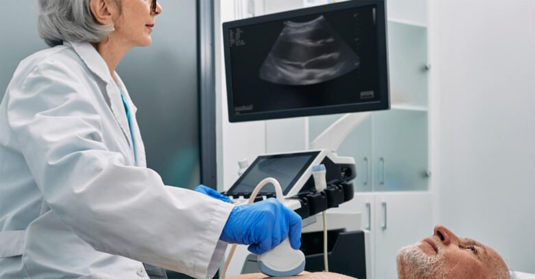
The tools we use to monitor and diagnose heart issues are crucial when aiming for better heart health. The use of cardiac ultrasound techniques changed the way in which the anatomy and function of the heart were viewed. These examinations are painless and are crucial to understanding the underlying cause of cardiovascular diseases and conditions so are an essential component of modern cardiology.
If you are about to undergo your first cardiac ultrasound examination, or if you simply want to know more about how these tests work—having this knowledge will help you strengthen your will to be more health-conscious. As new technologies allow us to take new images of the heart, let’s look at the field of cardiac ultrasound imaging—what they do, what their advantages are, and how they fit in the current medical world. Prepare yourself to learn so much more about this amazing area!
Indications for Cardiac Ultrasound Exams:
Ultrasound examinations of the heart can be categorized into several types based on the information required. The most frequently performed is transthoracic echocardiography (TTE). This interventional ultrasound-type exam is performed by an ultrasound technologist and echocardiographer.
Transesophageal echocardiography (TEE) can also be done. For better vision, a scope is placed in the esophagus. The views obtained by TEE can be quite exquisite and sometimes cannot be appreciated in TTE.
Stress echocardiography is a combination of ultrasound with exercise or medication that increases heart workload. The technique determines the capability of the heart to operate under stress.
Three-dimensional (3D) echocardiography provides improved visualization of the cardiac structures when compared to the 2D views provided by conventional echocardiography. Each exam serves a unique purpose in ensuring comprehensive cardiac evaluation and care.
Advantages of Cardiac Ultrasound:
For patients and healthcare practitioners, cardiac ultrasound also referred to as echocardiography, has a lot of advantages. All cardiac ultrasound methods are non-invasive, and fast, and allow real-time visualization of the heart anatomy and its function. This makes identifying and diagnosing various cardiovascular disease conditions much easier.
An important benefit is the evaluation of blood circulation in each of the heart chambers. Through these spaces, blood passes and doctors see how blood passes these areas, which enables diagnosis of other problems, for example, valve diseases or even some congenital problems.
On the contrary, another important advantage is safety in that there are no adverse effects due to the use of ultrasound waves. This technique does not involve X-ray or CT radiation and is thus appropriate for any human at any age.
Furthermore, cardiac ultrasound can be done at a faster rate, and in most cases, without the patient being admitted to the hospital. The results are often available on sight, and this helps in considerably faster decision-making on treatment strategies. This efficacy plus other factors translates into better patient care and also eases patient stress related to long waiting times.
How to Get Ready for a Cardiac Ultrasound Test?
The steps of preparing for a cardiac ultrasound exam are very easy and do not bring along complications or anxiety. First of all, dress appropriately so that there will be no need to alter your dress during the process. Any medications you may be on must be brought to your physician’s attention. Some medications might need to be suspended for a short time before the test. Walk into the facility after having some water; however, do not eat a heavy meal before eating. This way you augment the chances of clear images during the procedure.
If you have some anxiety concerning the process, feel free to ask for a family member or a friend for comfort. Their company is likely to positively affect how comfortable you are. When booking your appointment, remember to display any particular instructions as given by your health provider. Every center has individual rules that improve the encounter and its outcome.
The Significance of the Cardio-arts Images for the Patient:
One of the more challenging aspects of the exam is being able to interpret the results of a cardiac ultrasound. During the test, the images taken help in determining and providing information regarding heart structure and function. The achieved volume images of the heart assist in performing measurements such as the dimensions of the chambers, valves, and cardiac output. Each item provides different data about the heart status.
Normally functioning heart is expected to yield normal results without any anomalies. However, the increased size of chambers and valves not moving properly are some issues that signal potential underlying problems that require further probing. Your doctor will explain in detail what he or she has found out. If there’s something out of the ordinary that’s found, they may ask for further tests.
Being able to comprehend these results lets you make decisions in terms of your engagement in the treatment plan. This also helps you to pose questions and highlight concerns relating to where you are going with your heart health.
Common Uses and Applications of Cardiac Ultrasound:
- Another term for Cardiac ultrasound is echocardiography and this procedure has several critical applications in contemporary medicine. It is used mostly for the evaluation of the heart’s structure and functions. The physicians are able to see the chambers and valves of the heart and the tissues around the heart.
- This procedure is very useful for the diagnosis of heart murmurs as well as congenital heart disease. It aids Amol Egde in the detection of changes that would be difficult to see using other imaging techniques.
- Patients who have a past medical history of cardiovascular illness often receive this examination for follow-up purposes. With echocardiography, clinicians are able to snapshot images of structures in the heart and how those structures change in real-time.’
- A stress test with ultrasound looks at how well the heart works with physical activity. This technique helps determine the degree of physical activity that can be done and also some coronary artery problems.
- Furthermore, the importance of echocardiography is highlighted in the case of procedures where interventions are required, such as valve repairs or replacements, as it guides interventions and provides assistance appropriately. Its usage keeps on expanding as technology advances, which greatly improves patient care.
Advancements in Cardiac Ultrasound Technology:
The prospects of advancements in cardiac ultrasound technology are great, as improvements seem to be around the corner, which can further extend the diagnostic capabilities. AI is set to revolutionize image interpretation. It significantly improves the precision and turnaround time of algorithmic interpretations.
There is also increased popularity of portable devices. These small devices allow for scans to be conducted near the patients or in specific areas. With time, these technological advancements will increase the scope. Another area of interest is the scope of technology in telemedicine. Advanced imaging approaches that are used for ultrasound enable medical practitioners to assess heart disease without conducting a physical examination.
In addition, the advancement of 3D imaging has enabled the visualization of cardiac structures in greater detail. This development aids providers in making more accurate treatment plans that correspond to patients individually. As science evolves, so will new clinical contrast agents that can emphasize perfusion of blood flow and tissue more effectively and efficiently than what is already available on the market. The world of cardiac care is just about to change.
Conclusion:
Currently, echocardiographic methods are one of the most important contemporary methods of cardio diagnostics in clinical practice. They supplement the physician’s knowledge concerning the cardiovascular system. These painless exams are relevant for screening purposes. This technology promises to advance as well as further increase the accuracy of diagnosis.
The patient is at a great advantage considering the relative ease and ease of such tests. Knowing what will happen in advance can help ease the apprehension about such measures. As findings improve, we expect more changes that could enhance the results for patients. It is equally crucial for patients and healthcare providers to keep track of such changes. Better cardiac care means provisioning better heart health for all who are concerned.
FAQs:
1. What is a cardiac ultrasound?
An echocardiogram is done by sending ultrasonic waves and intercepting the waves after reflection as an ultrasonic echocardiogram. This test is performed to evaluate the structure and function of the heart in a reasonable and non-invasive manner.
2. Are there any risks associated with a cardiac ultrasound?
No, there are no noteworthy risks associated with this procedure. This is safe for most patients, including the pregnant female and a child.
3. How long does a majority of the cardiac ultrasound take?
The duration of most of these tests ranges from thirty minutes to sixty minutes, depending on the type of echocardiography being carried out.
4. Do you require any form of arrangement before your examination?
Preparation normally involves putting on rather loose garments in conjunction with not engaging in deep fasting so that one feels quite at ease during the activity.
5. When will my exam results be available?
Results can be tackled immediately after your exam or might take several days in case there are certain images that require further elaboration from the experts.


